CABG (Coronary Artery Bypass Graft)
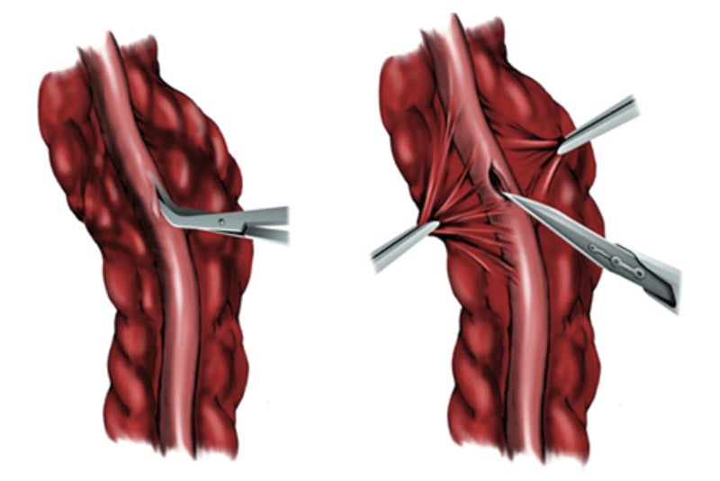
Arteriortomy
Arteriotomy should be proximal enough to offer the largest-sized coronary target, and just distal enough to avoid the area of obstruction.
Intramyocardial coronary branches often require sharp dissection of overlying tissue to identify the target site.
The arteriotomy is performed with a no. 11 blade and is then extended with fine. Pott’s scissors proximally and distally.
The arteriotomy should match the conduit diameter, and be at least 1.5 times the diameter of the distal coronary.
Distal anastomosis technique
A. An end-to-side anastomosis is accomplished with about 12 sutures.
B. Starting at 3 o’clock on the right side of the vessel, the suture is passed outside-in on the conduit and then inside-out at the corresponding location of the target vessel. Two more stitches are taken before the heel, one directly in the heel, and then two more on the left side of the anastomosis before parachuting the conduit down to the target vessel.
C. The anastomosis is then completed by placing another six stitches evenly spaced in the same manner for the duck beak.
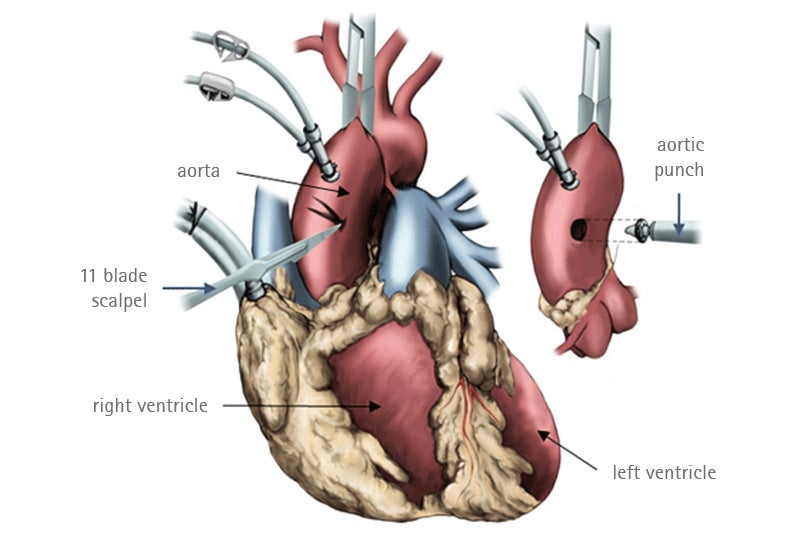
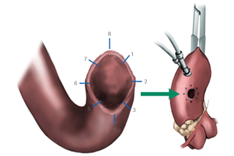
Proximal anastomosis technique
An arteriotomy is created with a no. 11 blade and a 4 mm aortic punch is used to create a circular aortotomy.
The anastomosis can be completed with 8-10 stitches in most cases; symmetry in the spacing between stitches is important to assure hemostasis.
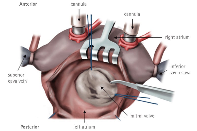
Valve Replacement
Excision of valve leaflets
A suture is placed into the anterior leaflet in order to displace it towards the surgeon.
A complete resection of the anterior leaflet is then performed.
Cases of extensive mitral valve stenosis may also require resection of the posterior leaflet.
Implant of the valve
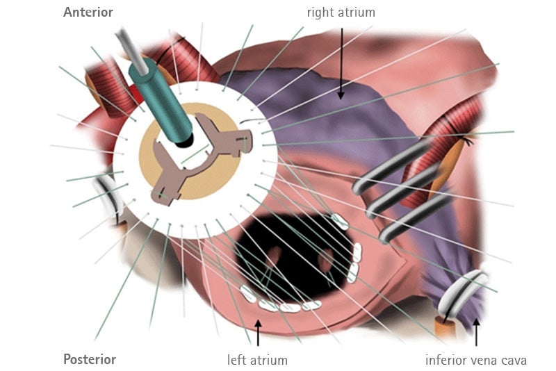
The annulus is sized to select the correct dimension of the valve.
Teflon reinforced sutures are placed into the sewing ring of the prosthetic valve after all annular sutures are placed.
Bioprosthesis insertion with gentle suture traction to avoid tangling.
Implant of the valve II
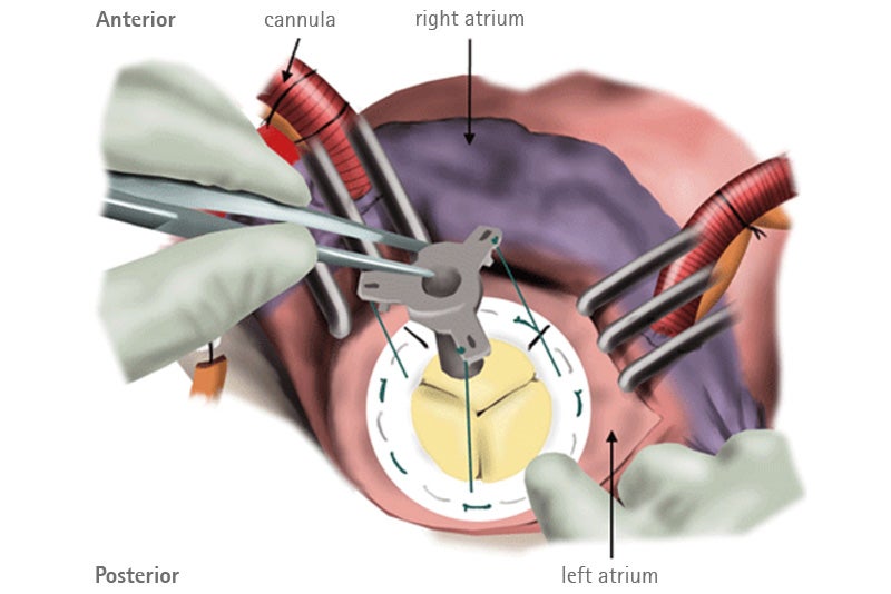
The prosthesis is slid into the annulus.
The holder is removed after all the sutures are tied while securing the bioprosthesis to the annulus.
The mechanical prosthesis is implanted in an anti-anatomic orientation and leaflet motion should always be checked.
Valve Repair
Annuloplasty
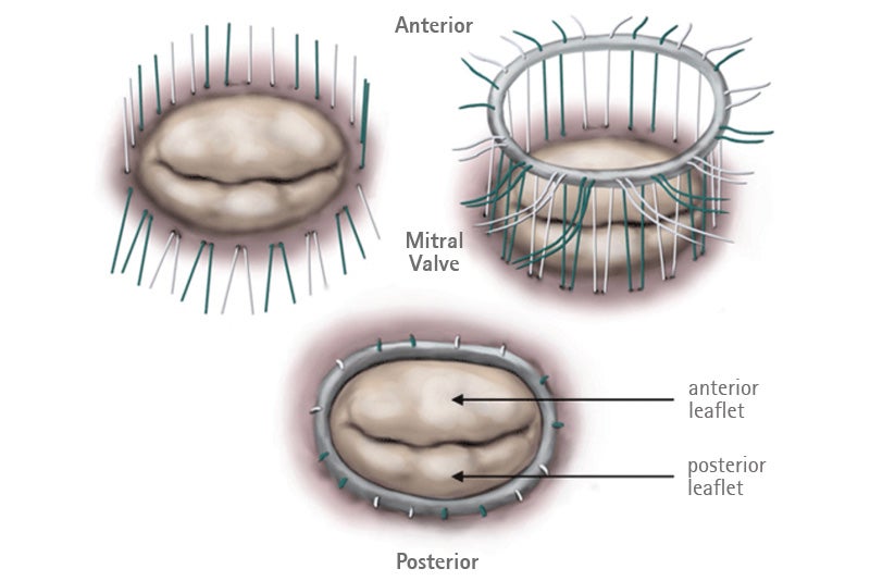
An annuloplasty device is usually used in all patients after mitral valve repair.
The first step is to determine the adequate size of the annuloplasty ring (between the two commisures).
Valve sutures are placed longitudinally through the mitral annulus.
Sutures are placed through the annuloplasty ring and then aproximated. The sutures are then knotted.
Mitral Posterior leaflet quadrangular resection
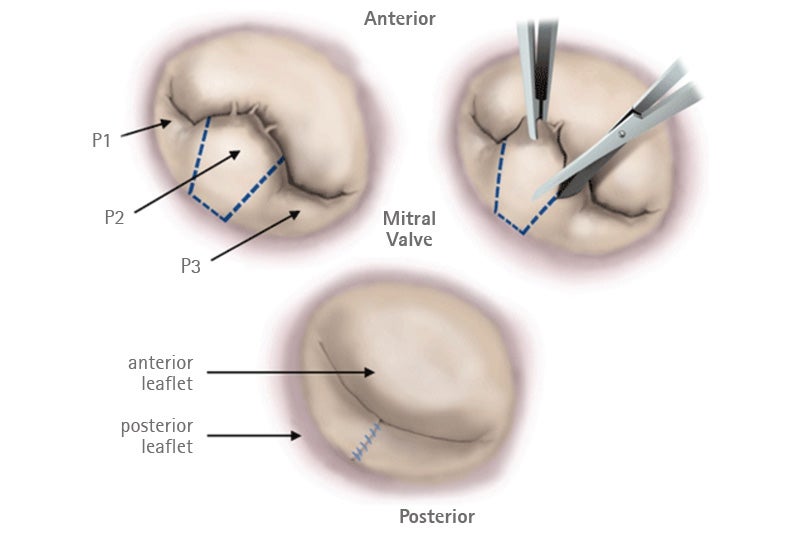
The most common defect that results in isolated valve incompetence is the mitral valve prolaps that typically involves the P2 leaflet.
Limited resection of this diseased section of leaflet (between the free margins of the P1 and P3 leaflets).
The remaining cleft between P1 and P3 is closed using interrupted sutures.
Sternal Closure
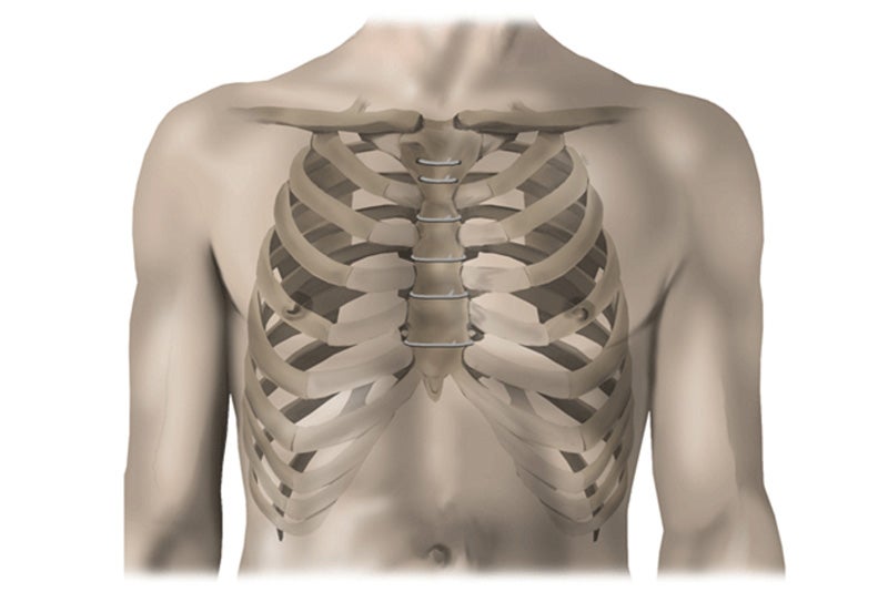
The sternum is reapproximated with six to eight stainless steel wires.
In the combined technique , the wires are passed around the sternum except in the case of the manubrium (upper part of the sternum), in which they are passed through the bone.
Care must be taken to avoid injury to the internal thoracic vessels.
